52 pounds of gravel, one Thoroughbred, and a connection that’s inspiring a future veterinarian

Jasper was on pasture rest for a knee injury. His owners had been carefully feeding him from a bucket, but the Thoroughbred gelding had a habit of knocking his feed over and eating it from the ground. Over time, however, he was getting more than his feed. He was also swallowing gravel. Fifty-two pounds of it.

“I’ve seen two severe gravel impactions causing complete obstructions, requiring surgery to resolve in the last six years,” said Carla Enriquez, clinical assistant professor of large animal emergency and critical care medicine at the Virginia-Maryland College of Veterinary Medicine. “It’s not an everyday thing.” 

On a summer morning, owner Gracelyn Bragg arrived at her North Carolina barn to find Jasper colicky. 

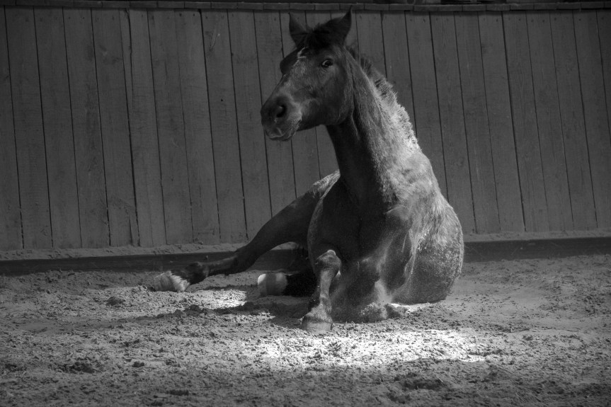
She walked him for hours — 44,000 steps, recorded on her phone — trying to keep him moving. A veterinarian came out, and for a while it seemed like they had turned a corner. Then Jasper went back in the pasture, went down, and wouldn’t get up. 

When they finally got him to his feet, they knew the window was closing. They loaded him on a trailer and headed to the Veterinary Teaching Hospital at Virginia Tech. 

Their field veterinarian had sedated Jasper heavily before loading him for the two-hour trip, and Enriquez had coordinated with her by phone the whole way. 

When Jasper stepped off the trailer, he immediately tried to lie back down. The team was ready. They had set up in the induction stall — where horses go when they may need to go straight to surgery — and worked him up there. 

“He was blowing through sedation,” Enriquez said. “He was very gassy. Reflux was coming out of his nose, which is not a good sign in horses. They can’t vomit, so when you see that, you know the pressure in his stomach is significant.” 

The blood work came back clean. He was a good surgical candidate. Within a little over an hour of arriving, Jasper was on the table. 
Read the full story…

Horse emergency? Be ready to haul

Large animal veterinarians are already in short supply, and for horse owners in our region, that crunch just got a little tighter.

If you rely on Equine Field Services through Virginia Tech, there’s an important update to how after-hours emergencies will be handled. Most after-hours equine emergencies will now require hauling your horse to the Veterinary Teaching Hospital.

What’s changing?

To improve response time and care, all emergency calls after 4 p.m., as well as weekends and holidays, will be handled at the hospital rather than on the farm.

That means if your horse has an emergency, you’ll be asked to bring them directly to the VTH, where a full team and hospital resources are immediately available.

The emergency fee for hauling in will be $80 for Equine Field Services clients.

Why the shift?

This change is coming from a few realities many of us in the horse world are already feeling:

  • Fewer field service veterinarians available.
  • A growing number of emergency calls.
  • The need to get horses seen and treated as quickly as possible.

By centralizing after-hours care at the hospital, the goal is to:

  • Reduce wait times in critical situations.
  • Provide faster access to diagnostics and treatment.
  • Support the sustainability and well-being of the veterinary team.

What if you can’t haul?

They understand that hauling isn’t always possible.

If a veterinarian determines that your horse cannot be safely transported, on-farm emergency care will still be available. However, there is an increased cost:

  • On-farm ER fee: $175
  • (This is in addition to standard emergency and trip fees)

What this means for horse owners

This is one of those changes that may take a little adjustment, especially if you’re used to farm calls for emergencies. But in many cases, getting your horse to a hospital setting faster can make a real difference in outcomes.

It’s also a good reminder to:

  • Have a trailer plan in place.
  • Know who you can call to help haul in an emergency.
  • Keep emergency contacts easily accessible.

New table for CT scanner allows examination of horses at Veterinary Teaching Hospital

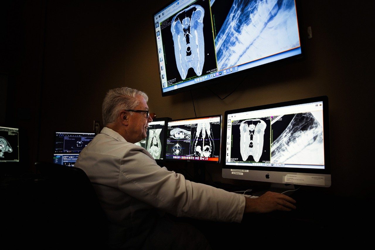
Gregory Daniel, professor of radiology in the Department of Small Animal Clinical Sciences at the Virginia-Maryland College of Veterinary Medicine in Blacksburg. Photo by Andrew Mann for Virginia Tech.
By Virginia Tech

Until recently, clinicians at the Veterinary Teaching Hospital in the Virginia-Maryland College of Veterinary Medicine couldn’t use CT, or computed tomography, for large animals. But the recent purchase of an $85,000 table for large animals that can be wheeled to the CT scanner now allows for that option, particularly useful for studying conditions inside the heads of equine animals.

The Veterinary Teaching Hospital in Blacksburg will be the only veterinary hospital offering the capability to employ CT scans for large animals in a radius extending roughly 240 miles, to as far away as North Carolina State University’s veterinary college in Raleigh and to the Marion duPont Scott Equine Medical Center (EMC) in Leesburg, where there is a CT scanner that allows equine animals to stand during the exam.

“We have referred some of these cases to the EMC because we needed the additional information and that was the best thing for the patient,” said Chris Byron, head of the Department of Large Animal Clinical Sciences. “If they’re coming from southern Virginia or North Carolina to us, it’s a big ask to send them another 3 1/2 to four hours north. So for our region, for Southwest Virginia and into West Virginia, it’s certainly a big, big step up in capabilities.”

“We commonly use the CT scanner for imaging dogs and cats,” said Gregory Daniel, professor of radiology in the Department of Small Animal Clinical Sciences. “The current CT scanner works great for small animals, but most scanners have a 450-pound table limit. For most people and certainly for all small animals, that is sufficient. But if you’ve got a 1,000-pound patient, then a traditional CT scanner cannot accommodate that weight.”

Now, a horse or other large animal can be anesthetized, laid upon the table, and rolled to the CT scanner. The table is electronically synced with the scanner, allowing it to capture slice-by-slice imagery of a large animal’s skull, teeth, and cranial cavities, providing much more detail than X-ray radiographs can reveal.

Byron and Daniel both describe the acquisition of the CT scanner table for large animals as a “game-changer.” Combined with MRI technology that does well with ligaments and cartilage of large animals’ legs and feet, plus existing radiograph and ultrasound abilities, veterinary clinicians can cover much more of a large animal’s body in examinations.

“Radiographs underestimate the extent of the disease,” Daniel said. “But the CT scanner can greatly improve our ability to diagnose diseases of the head and nasal passages.”

Read more.